
Every step of our development is guided by real-world surgeon feedback, current clinical practice, and peer-reviewed scientific evidence.

OrthoPlanner™ improves surgical outcomes through precise 3D preoperative planning, reducing intraoperative ambiguity and improving fixation accuracy. The following resources summarize our clinical publications, case studies, and outcome data.
Enter your email to receive our comprehensive clinical evidence summary — covering published outcomes, case studies, and validation data.

reduction in OR time
fixation accuracy
surgeons more confident
See related: Procedures · OrthoPlanner™
surgeons more confident
Downloadable case study PDFs.
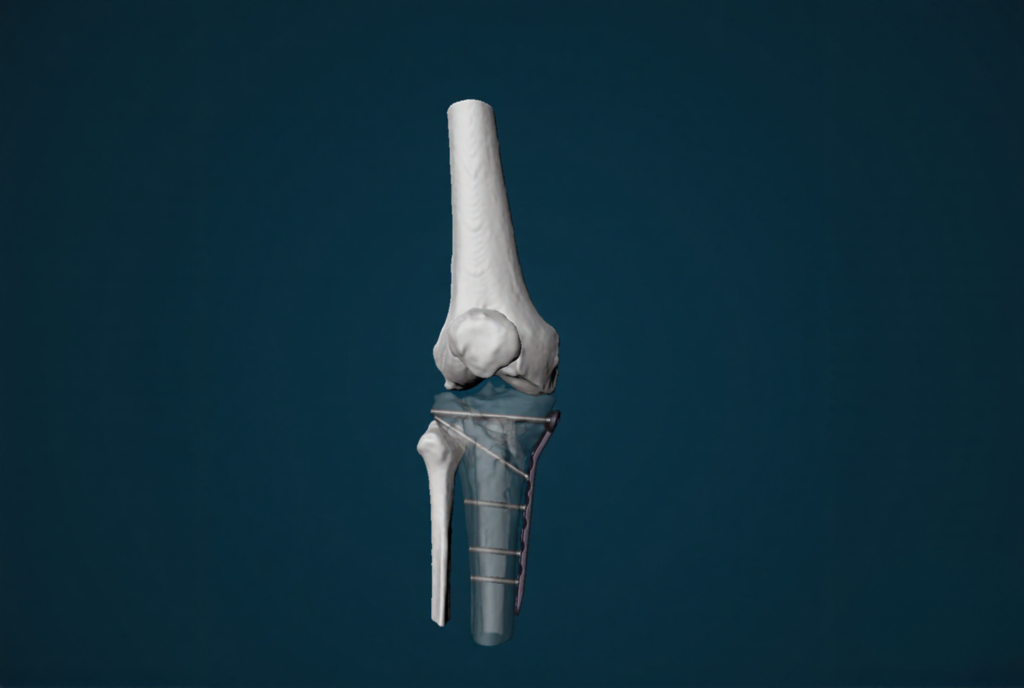
Dr. Arvind von Keudell · Harvard Medical School & University of Copenhagen
A rare flexion–varus–internal-rotation tibial plateau injury in a 68-year-old that first presented as a classic Schatzker IV. 3D planning exposed the rotational component invisible on X-ray and CT, clarifying the reduction vectors and confirming a directly medial buttress-plate position among several candidates. Anatomic reduction was achieved only once the rotational correction was applied — restoring coronal alignment and a congruent joint line.
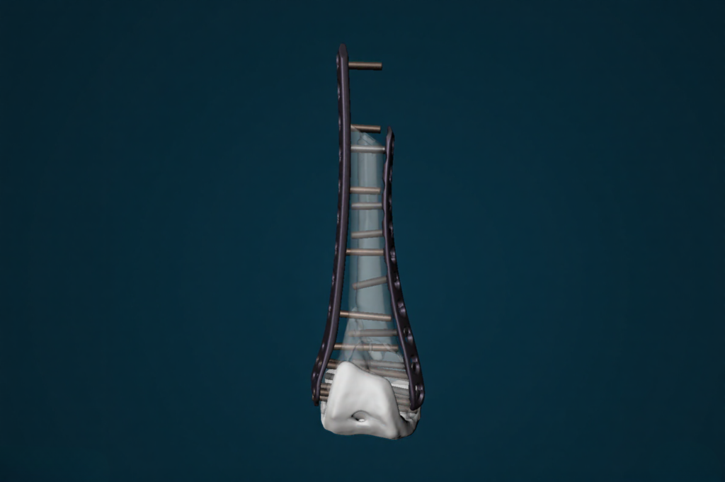
Prof. Benedikt Braun · BG Klinik Tübingen
A comminuted supracondylar fracture (Lewis–Rorabeck II) above a well-fixed knee replacement in a 75-year-old. Preoperative segmentation and virtual reduction established a dual medial-and-lateral plating strategy, with screw lengths and periprosthetic trajectories defined before incision. The construct matched the plan intraoperatively and was stable enough to permit immediate, unrestricted weight-bearing.
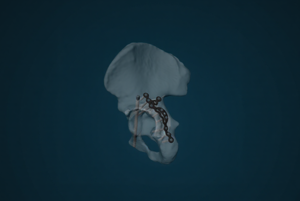
Prof. H. Claude Sagi · University of Cincinnati Medical Center
A transtectal transverse-plus-posterior-wall acetabular fracture (Letournel) in a 46-year-old following a high-energy motor vehicle crash. Virtual reduction defined the anterior-column lag-screw corridor and the ITS buttress-plate placement ahead of a Kocher–Langenbeck approach. The final construct agreed with the preoperative plan, and the fracture healed without loss of reduction or avascular necrosis at three months.
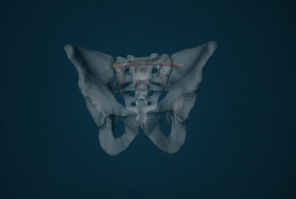
Prof. H. Claude Sagi · University of Cincinnati Medical Center
A U-type sacral fragility fracture in a 71-year-old with a sacralized L5 and dysmorphic first sacral segment — anatomy that would not accept a straight implant. Planning mapped the curved osseous corridor for a flexible trans-sacral–trans-iliac implant, enabling a fully percutaneous fixation. Follow-up radiographs confirmed maintained alignment with no implant failure.
Enter your details to unlock the full library of documented cases.
The science behind OrthoPlanner™, published in leading journals.